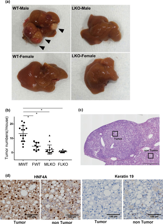
Figure 7.
NASH-induced liver tumours in Bcl6-LKO mice (a) WT and Bcl6-LKO mice were fed with CDAHFD for 38 weeks. Macro images of liver tumours induced are shown. (b) The number of liver tumours induced by 38 weeks CDAHFD feeding was counted. Results are represented as mean ± standard deviation (S.D.) (n = 15 for male wild-type mice, n = 8 for female wild-type mice, n = 17 for male Bcl6-LKO mice, n = 11 for female Bcl6-LKO mice). *P < 0.05. MWT, male wild-type mouse samples; FWT, female wild-type mouse samples; MLKO, male Bcl6-LKO mouse samples; FLKO, female Bcl6-LKO mouse samples. (c,d) Analysis of liver tumours induced by long-term CDAHFD feeding. Haematoxylin and eosin staining and immunohistochemical staining were performed. Representative images are shown. The scale bar shows 1 mm in (c) and 100 μm in (d).

