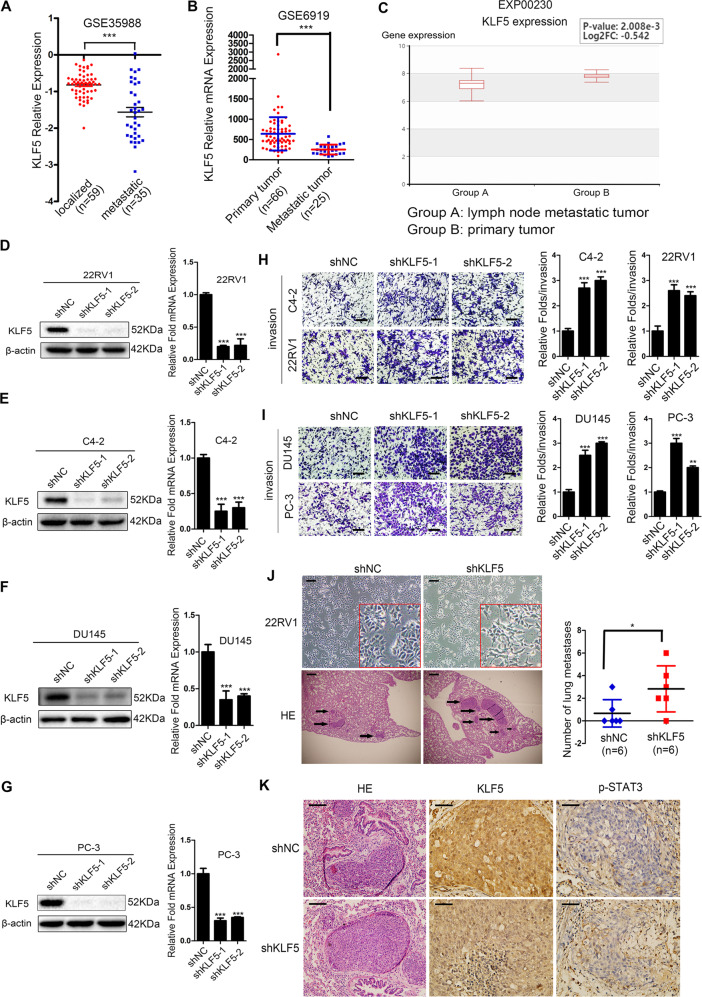
Fig. 2. Knockdown of KLF5 promotes invasion of prostate cancer cells in vitro and in vivo.
a, b KLF5 mRNA expression in localized and metastatic tissues of prostate cancer from GEO databases GSE35988 and GSE6919. c KLF5 mRNA expression was lower in lymph node metastatic tumors than in primary prostate tumors as per EXP00230 of the HCMDB website. d–g Real-time quantitative PCR and western blotting of KLF5 mRNA and protein levels in C4-2, 22RV1, DU145, and PC-3 cells transfected with KLF5 shRNA (shKLF5) or negative control (NC). 18S and β-actin were used as loading controls. h, i Representative Transwell data and quantification analysis of invasion assays in C4-2, 22RV1, DU145, and PC-3 cells transfected with KLF5 shRNA or shNC. Scale bar = 100 μm. The data are from at least three independent experiments expressed as the mean ± SD. *p < 0.05; **p < 0.01; ***p < 0.001. J Representative microscopic images of the morphology of 22RV1 cells after KLF5 knockdown (upper panel), and representative histological (lower panel) and quantification analyses of nude mice lung metastatic foci in 22RV1/NC and 22RV1/shKLF5 groups. Scale bar = 100 μm. k Immunohistochemistry staining of KLF5 and p-STAT3 in nude mice lung metastatic tissues from 22RV1/NC and 22RV1/shKLF5 groups. Scale bar = 100 μm.

