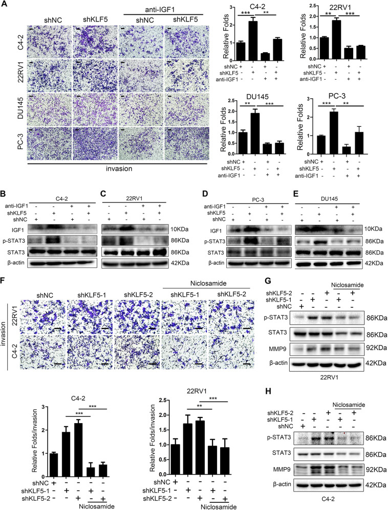
Fig. 5. Blocking of IGF1/STAT3 pathway suppresses the cell invasive ability enhanced by KLF5 downregulation.
a Representative Transwell invasion photographs and quantification of invasive abilities of shKLF5 subtypes of C4-2, 22RV1, PC-3, and DU145 cells treated with neutralizing antibody of IGF1 for 24 h. **p <0.01, ***p <0.001 versus control. Scale bar = 100 μm. b–e Western blotting analysis of IGF1, p-STAT3, and STAT3 expression in C4-2, 22RV1, PC-3, and DU145 cells transfected with shKLF5 or shNC after treatment with neutralizing antibody of IGF1 for 24 h. f Representative pictures and quantification analysis of invasion assays in C4-2 and 22RV1 cells transfected with shKLF5 or shNC after treatment with STAT3 inhibitor niclosamide. Scale bar = 100 μm. Each experiment was repeated at least three times and the result of a representative experiment is shown. **p < 0.01, ***p < 0.001. 18S was used as an internal loading control. g, h Western blotting analysis of KLF5, p-STAT3, STAT3, and MMP9 expression levels in C4-2/shKLF5 (g) and 22RV1/shKLF5 (h) sublines after treatment with 0.5 μM of STAT3 inhibitor niclosamide for 24 h.

