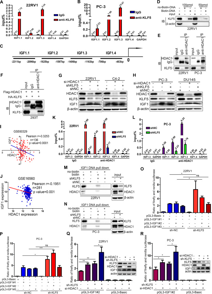
Fig. 6. KLF5 binds on the IGF1 promoter cooperatively with HDAC1 and suppresses the transcription of IGF1.
a, b Chromatin immunoprecipitation (ChIP) assay of binding sites of KLF5 on the IGF1 promoter detected by qPCR in 22RV1 and PC-3 cells. GADPH promoter was served as a negative control. c The map of IGF1 promoter, which indicates the regions analyzed in ChIP assay. d Oligonucleotide DNA pull-down assay was performed to verify that the DNA fragment amplified by PCR could pull-down KLF5 protein in 22RV1 cells; the pull-down protein was detected by western blotting. e, f Co-immunoprecipitation (Co-IP) assay of the mutual interaction of KLF5 and HDAC1 proteins at endogenous (22RV1 and PC-3) (e) and exogenous levels (293T) (f), as detected by western blotting analysis. IgG was used as the negative control, while input was a positive control. g, h Western blotting analysis of HDAC1, KLF5, and IGF1 protein expression in 22RV1, C4-2, PC-3, and DU145 cells transfected with shKLF5 lentivirus and si-HDAC1 siRNA. i, j Expression of IGF1 and HDAC1 mRNA was negatively correlated in prostate cancer tissues, as per analysis in GSE16560 and GSE60329. k, l ChIP-qPCR assay of HDAC1 binding on IGF1 promoter in 22RV1 and PC-3 cells transfected with shKLF5 or shNC. Treatment with IgG was used as a negative control. m, n Oligonucleotide DNA pull-down assays were performed to verify that 100 pmol specific DNA fragment precipitated HDAC1 protein in NC and shKLF5 sublines of 22RV1 and PC-3 cells. No-biotin labeled DNA was used as a negative control. o, p Dual-glo luciferase assay indicated that knockdown of KLF5 in PC-3 and 22RV1 cells promoted the transcriptional activity of IGF1 promoter. q, r Dual-glo luciferase assay showed that knockdown of HDAC1 further increased the transcriptional activity of the IGF1 promoter activated by knockdown of KLF5 in 22RV1 and PC-3 cells. ns p > 0.05; *p <0.05, **p <0.01, ***p <0.001. The pGL3-IGF1#1, pGL3-IGF1#2, and pGL3-IGF1#3 indicate three different oligonucleotides located in the proximal region of the IGF1 promoter. Plasmid pGL3-Basic served as a negative control. KLF5, HDAC1, and IGF1 expression in 22RV1 and PC-3 cells treated with shKLF5 and si-HDAC1 detected by western blotting.

