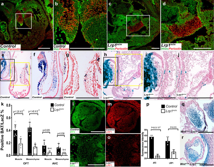
Fig. 5. Abnormal atrioventricular and outflow tract cushion development was observed in Lrp1m/m mutants.
Immunostaining of NFATC-1 (green) and Periostin (red) in wildtype (a, b) and Lrp1m/m mutant (c, d) embryos at E12.5. b, d Magnified views of the white box from (a, c). Compared with the well-formed two atrioventricular valves in the wildtype control, Lrp1m/m mutant demonstrated primitive undivided endocardial cushion morphology. Representative images from X-gal and Eosin stained tissue sections from E10.5 control (Lrp1m/+) (e–g) and Lrp1m/m mutant (h–j). f, i The high magnified view of the black box e and h (OFT). g, j The high magnified view of the yellow box from (e, h) (AV cushion). X-gal staining e–j demonstrated diminished expression of LacZ expression in the Lrp1m/m AVC (e, f, h, i) and OFT cushion (e, g, h, j). k Quantitative analysis of the percentage of positive BAT/LacZ cells in the muscle and mesenchyme portion of OFT and AVC. Lrp1m/m mutant had hypocellular AVC and OFT cushions (h–j). l–o Immunostaining of AP2α (green) and LRP1 (red) in Lrp1m/m mutant (n, o) and control (l, m). Lrp1m/m mutant had decreased expression of AP2α and LRP1 in the outflow tract at E10.5–E11.5 (l–o). p Quantitative analysis of the fluorescence intensity of AP2α and LRP1 demonstrated decreased expression of both AP2α and LRP1 in Lrp1m/m mutant OFT. (q, r) LacZ staining of OFT in Wnt1+/cre: Lrp1f/f/rosa/rosa and control (Wnt1+/cre: Lrp1f/+/rosa/rosa), which demonstrated decreased LacZ expression in the Wnt1+/cre: Lrp1f/f/rosa/rosa mutant. Scale bars: 200 μm. Statistical comparison was performed using unpaired two-way Student's t test. Error bars show standard deviation.

