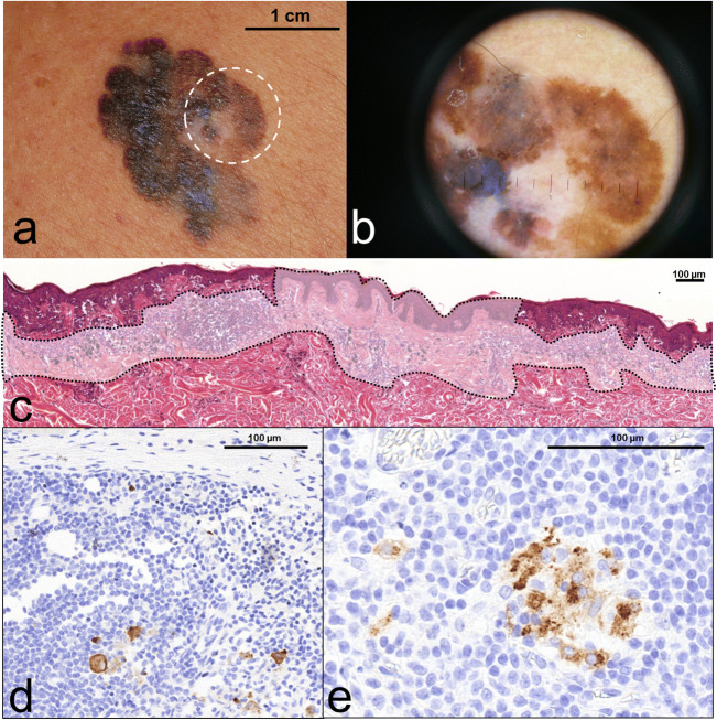
Fig. 1.
shows a case report of a male patient aged 71, presenting regressing superficial spreading melanoma on his back region (a). At dermoscopy, the centre of the polychrome plaque displayed greyish-whitish area with peppering sign which is characteristic for regression (b). After the surgical removal of tumour, the histopathology showed extensive vanishing of junctional and dermal melanoma cells replaced by fibrosis, accumulation of melanophages and lymphocytic infiltrate together with focal neovascularisation (c - circumscribed faded area). Only the edges of the presented section contained atypical residual microinvasive melanoma cells within the regressive microenvironment. Although calculated Breslow thickness from the residual melanoma counterpart showed only 0.532 mm, dermal mitotic activity together with the adverse regression indicated SLNB. During the histopathological processing right axillary SLN contained scattered metastatic melanoma cells (d) which were also present in two other lymph nodes in the right axillary dissection sample (e)

