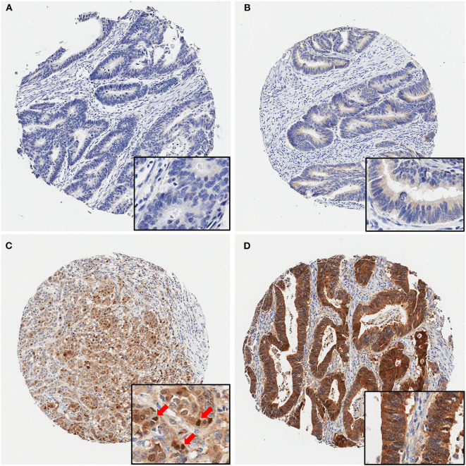
Figure 2.
Representative immunohistochemical maspin protein stains showing four different TMA cores. (A) Negative (no protein expression), (B) weakly positive, (C) moderately positive (with several positive nuclei, red arrows), (D) strongly positive staining. Original magnifications: 40× (insets, 400×).

