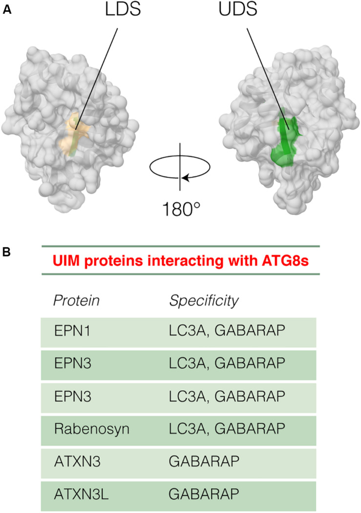
FIGURE 7.
Interaction between UIM motifs and ATG8 proteins. (A) The figure illustrates the location on the 3D structure of the yeast ATG8 protein of the LIR Docking Site (LDS, tan) and the UIM Docking Site (USD, green) using the PDB entry 3VXW (Kondo-Okamoto et al., 2012). (B) The table shows the UIM-containing proteins that bind LC3A or GABARAP with respective specificities.

