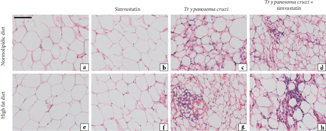
Figure 5.

Representative photomicrographs of histological sections of adipose tissue from uninfected and infected C57BL/6 mice with T. cruzi (VL-10 strain). The acute inflammatory process was evaluated in the adipose tissue of mice infected with T. cruzi and fed with high-fat diet, under simvastatin treatment or not, as indicated in the figure. Tissues were stained with hematoxylin and eosin (H&E) at 30 days after infection. Scale bar = 50 mm.
