Figure 1.
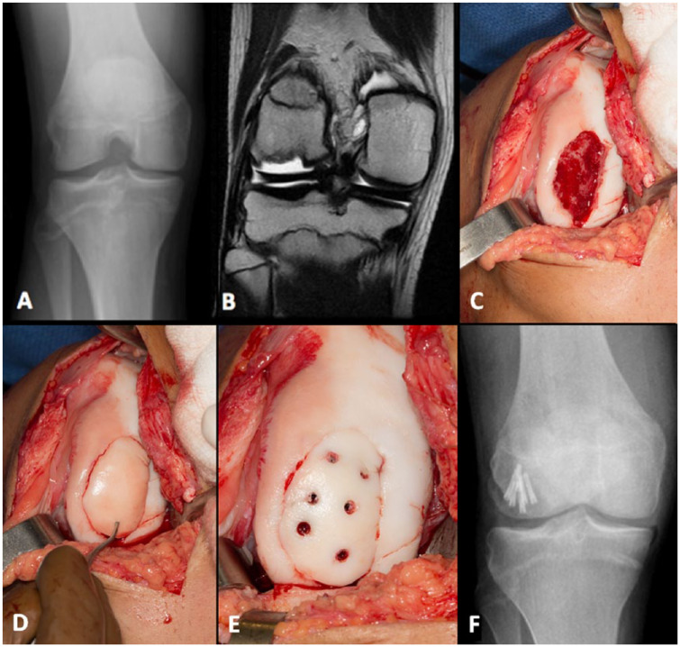
Preoperative (A) radiograph and (B) magnetic resonance image of a large lateral femoral condyle lesion with subsequent (C) intraoperative lesion bed visualization and preparation, (D) fragment reduction, and (E) fixation. (F) Postoperative radiograph demonstrating fragment fixation. Patient subsequently returned to competitive basketball with no pain.

