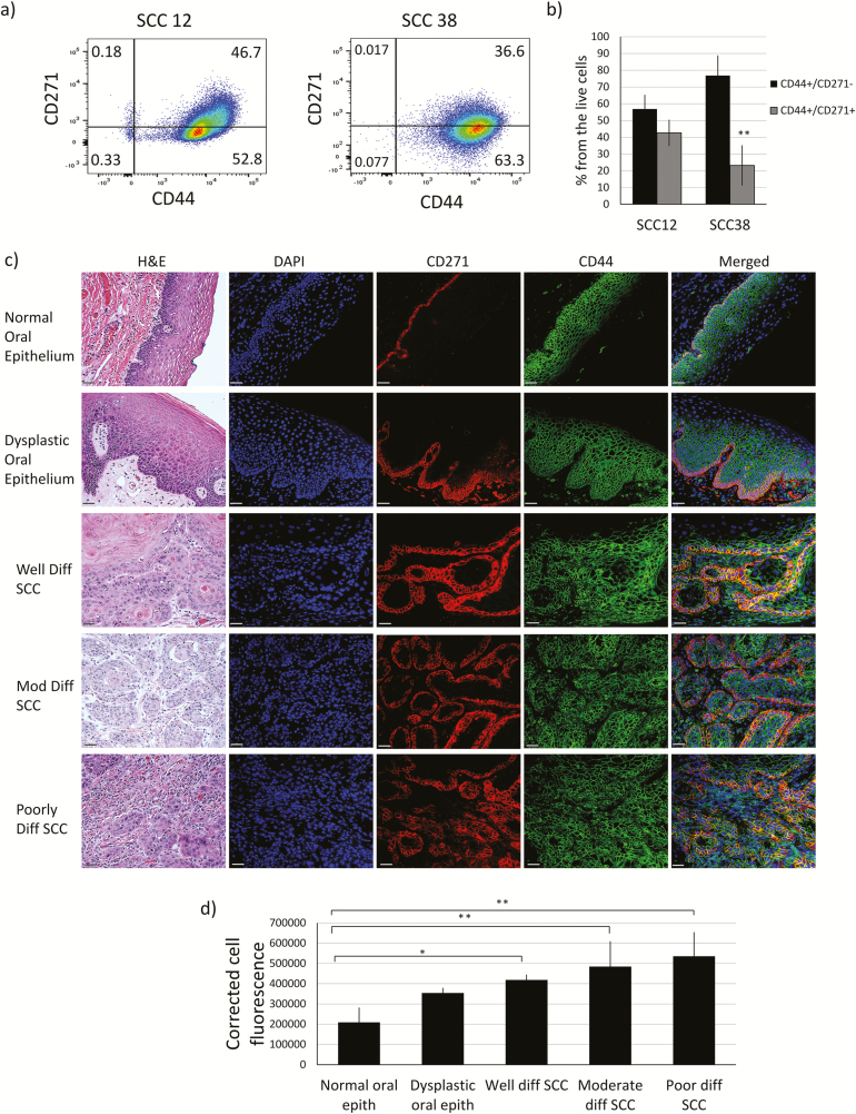
Figure 1.
CD271+ cells are a subpopulation of CD44+ cells. (a) SCC12 and SCC38 cells were examined for the expression of CD44 and CD271 markers using flow cytometry. (b) Percentage of CD44+/CD271− cells and CD44+/CD271+ cells in SCC12 and SCC38. (c) Human normal and oral SCC samples were stained with H&E and monoclonal immunofluorescence antibodies against CD44 and CD271 for the assessment of the tumor grade and the localization of CD44+ and CD271+ cells (×20 magnification. Scale bar: 37 μm). (d) Quantification of cell fluorescence of CD271+ from stained images in (c) with ImageJ software (n = 3 to 6 per group). Data are presented as mean ± SD (*P < 0.05 and **P < 0.01). ).

