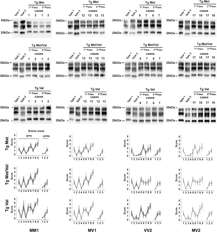
FIG 1.
PrPres Western blot profiles (top) and vacuolar lesion profiles (bottom) in the brains of mice expressing human PrP inoculated with sporadic CJD (sCJD). y score indicates intensity of vacuolar lesions; x axis indicates standard brain gray areas (1 to 9) and white areas (1 to 3). Transgenic mice that express the Met129 (tgMet), Val129 (tgVal) human PrP and their crossbreed (tgMet/Val) were inoculated intracerebrally (6 mice, 20 μl per mouse) with a 10% brain homogenate (frontal cortex) from sCJD cases that were Met129 (MM) homozygotes, Val129 (VV) homozygotes, or Met/VAl129 (MV) heterozygotes and displayed either a pure type 1 or a pure type 2 PK-resistant PrP (PrPres) Western blot (WB) isoform (Table 1). Two iterative passages were performed for each line (Table 1). After the second passage and in each mouse line, the isoform (type 1/type 2) of the PrPres was determined in the mouse brain by sodium dodecyl sulfate-polyacrylamide gel electrophoresis (SDS-PAGE) and WB with the anti-PrP monoclonal antibody Sha31 (epitope YEDRYYRE). A PrPres type 1 isoform (MM1 sCJD isolate) and type 2 isoform (VV2 sCJD isolate) were included as controls on each gel. The WB results obtained in each line are reported in Table 1. In parallel, standardized vacuolar lesion profiles were established in the brains of the same mice. Symbols: in the MM1 lesion profile graphs, ▽, case 1; ▵, case 2; ○, case 3; ◊, case 4; □, case 5; in the MV1 lesion profile graphs, ▵, case 12; ○, case 13; □, case 14; in the VV2 lesion profile graphs, ▽, case 6; ▵, case 7; ○, case 8; ◊, case 9; □, case 10; in the MV2 lesion profile graphs, ○, case 17; □, case 18.

