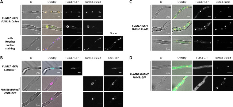
FIG 5.
Fum3, Fum8, Fum17, Fum18, and Cer1 localization in F. verticillioides. Confocal microscopy for localization of Fum17-GFP and Fum18-DsRed (A), Cer1-BFP (B), DsRed-Fum8 (C), and Fum3-GFP (D). Conidia were inoculated in ICI/6 mM Gln and grown as a standing culture overnight. The indicated double mutants were analyzed for GFP, DsRed, and/or BFP fluorescence. The cells were untreated, except when indicated (nuclear staining with Hoechst 33342 in panel A). Shown are individual channels in black/white, bright-field (BF) images and an overlay with the BF in color.

