
Fig. 7.
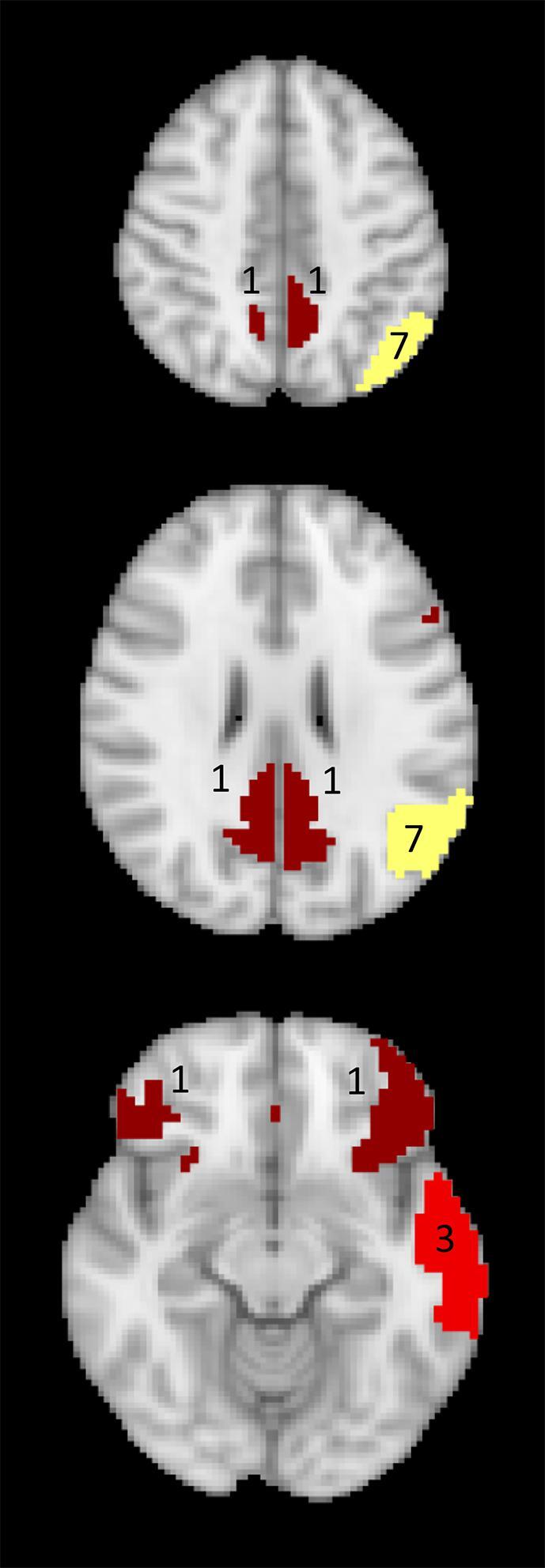
Mapping of the number of neurocognitive tests (out of 10) that significantly correlated with the connectivity of the DMN nodes in glioma patients. Red: left lateral temporal cortex DMN node, yellow: left inferior parietal lobule DMN node, dark red: bilateral mPFC nodes and PCC nodes.