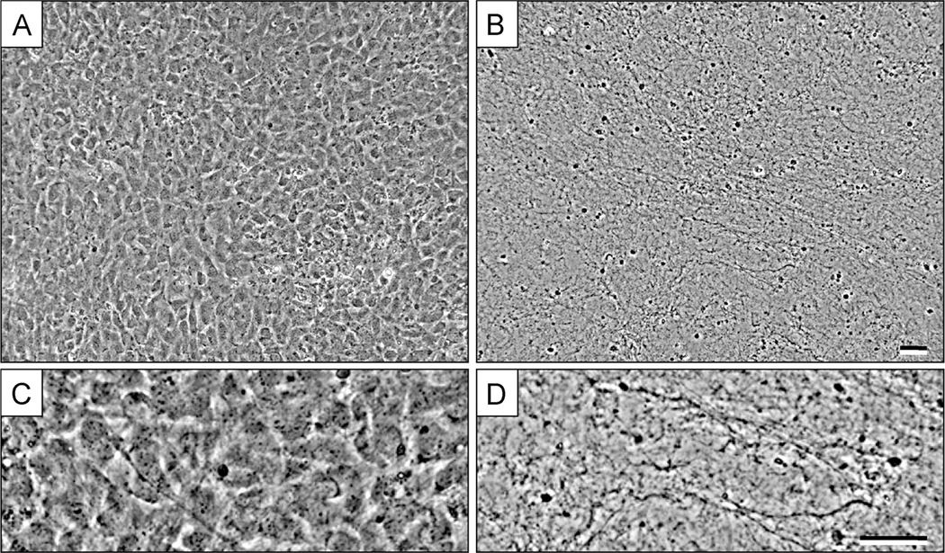
Figure 3. Fibroblastic cell-derived 3D ECM (fCDM) before and after extraction process.
A, shows a representative image of fibroblasts (NIH-3T3) in 3D culture at day 5, prior to matrix extraction. B, show the resulting 3D fCDM fibers after the decellularization process. Panels C and D are magnified areas from images A and B, respectively. Note the fibrous pattern and the absence of cell bodies and debris in D. Bars represent 50 μm. Image was adapted from Cukierman E., Preparation of Extracellular Matrices Produced by Cultured Fibroblasts. Curr Protoc Cell Biol, 2002 (Edna Cukierman, 2002). Copyright © 2002 by John Wiley & Sons, Inc.

