Figure 7.

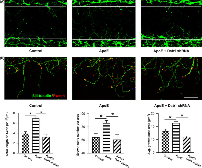
ApoE improves axonal outgrowth and growth cone formation in injured neurons. Axons were stained at 24 h after mechanical injury. (A) Representative images of axonal outgrowth after scratch injury. βIII‐tubulin‐positive neurites (green) that crossed over the white line margin were measured to quantify the regenerated axonal length. Scale bar = 200 µm. ApoE treatment induced significant elongation in total axonal length compared to vehicle control treatment. However, compared to the ApoE treatment group, the Dab1 shRNA pretreatment group exhibited an attenuation of total regenerated axonal length due to Dab1 expression downregulation (P < .05). (B) Representative images of growth cone regeneration. Phalloidin staining (red) showed growth cones at the tips of regenerating βIII‐tubulin‐positive neurites (green). Scale bar = 50 µm. The number of growth cones visible per area in the lesion gap and the average growth cone area were quantified. Along with the regenerated axonal extension, the number of growth cones was significantly higher after ApoE treatment than the number in the control group. Likewise, the average area per growth cone in the ApoE‐treated group was significantly larger than that in the control group (P < .05). However, the effects of ApoE were abolished when Dab1 was downregulated by pretreatment with Dab1 shRNA. *P < .05. n = 6/group
