
FIGURE 1.
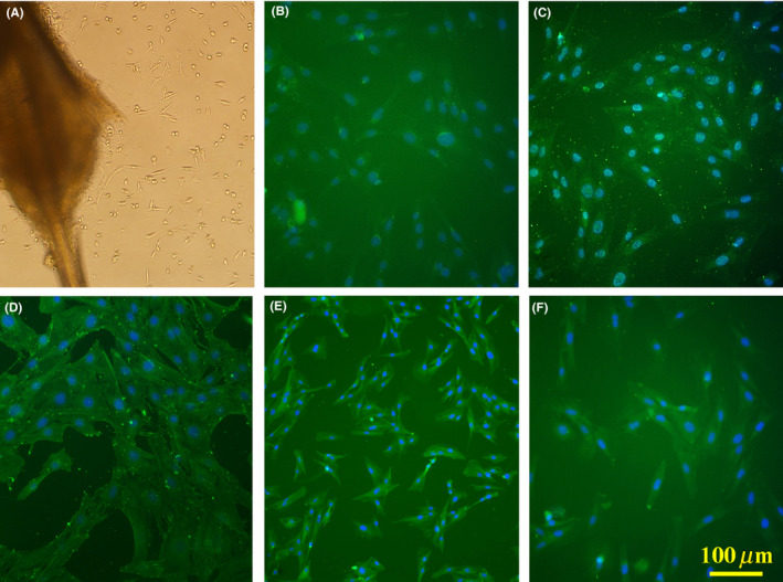
Migrated cells around the bulge, 7 d after explantation (A). Immunostaining against nestin (B), SOX10 (C), doublecortin (D), β‐III tubulin (E), and glial fibrillary acidic protein (F) to verify migrated cells. Cell nuclei counterstained with DAPI. Scale bar: 100 μm