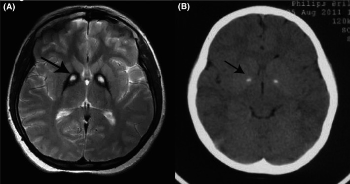
Figure 1.

Neuroimaging of patients with PKAN. A, T2‐weighted MRI of the brain showed a specific pattern of hyperintensity (indicated by the arrow) within the hypointense medial globus pallidus (“eye‐of‐the‐tiger” sign). B, Brain CT showed calcification in medial globus pallidus, which was indicated by the arrow
