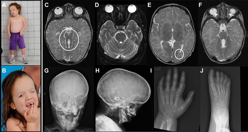
FIG. 1.
Clinical photographs and images of the patient. (A) Patient had severe growth retardation associated with relative macrocephaly. (B) Craniofacial features included tall forehead, widely spaced eyes, epicanthal folds, severe ptosis of left eye, and borderline low set ears. Brain MRI at 2 months showed molar tooth sign (circle) (C) in comparison to normal (D), left occipital cortical heterotopia (circle) (E), and hypoplastic extraocular muscles (F). Skull X-rays showed a large head with increased craniofacial ratio, copper beaten marks, and frontal bossing (G and H). Abnormal cone-shaped epiphyses were noted in hand (I) and feet (J) X-rays.

