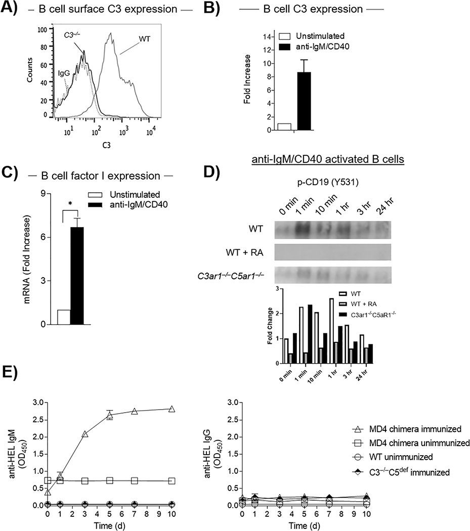
Figure 5. B2 cells endogenously phosphorylate CD19 by virtue of locally producing C3 and factor I.
A) WT B cells were adoptively transferred into C3−/− mice (n=5) after which C3 expression on B cells was assayed by flow cytometry. B) WT splenic B2 cells were incubated at 37°C for 72 h with anti-IgM F(ab’)2/anti-CD40, after which C3 mRNA expression was quantified by qPCR C) WT splenic B2 cells were incubated as in B) after which factor I mRNA levels were quantitated by qPCR. D) WT B2 cells in the absence or presence of RA (100 ng/mL each) or C3ar1−/−C5ar1−/− B2 cells were incubated at 37°C with anti-IgM F(ab’)2/anti-CD40 (3 μg/mL) and CD19 phosphorylation assayed on immunoblots as a function of time. E) MD4 BM was transplanted into irradiated C3−/−C5nul recipients. Following immunization of the chimeras and the untreated C3−/−C5nul mice with HEL in CFA, blood samples were drawn at 1, 3, 5, 7 and 10 d and assayed for by ELISAs for anti HEL IgM (left) and IgG (right) Abs. As controls, blood samples were assayed from identically immunized WT mice and from unimmunized C3−/−C5nul-MD4 chimeras.

