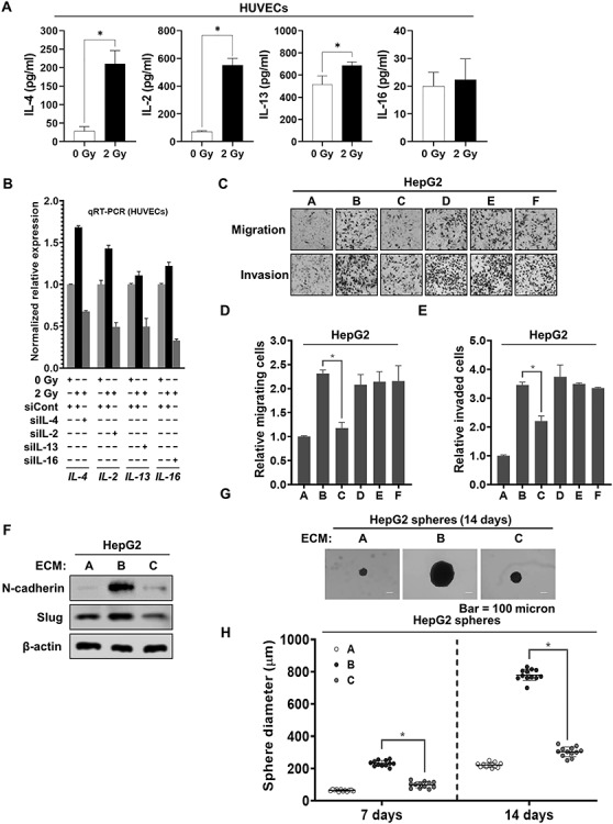
Fig. 1.

IL-4 released from 2 Gy–irradiated endothelial cells contributed to increasing the malignancy of liver cancer cells. (a) The secretion of IL-4, IL-2, IL-13 or IL-16 in 2 Gy–irradiated HUVECs. After HUVECs were irradiated with 2 Gy, and then cultured for 24 h, ELISA was performed to detect the secretion of interleukins, using conditioned medium obtained from HUVECs. (b) qRT-PCR analysis of the mRNA expression levels of IL-4, IL-2, IL-13 and IL-16 in the presence or absence of siRNAs targeting IL-4, IL-2, IL-13 and IL-16, respectively, in 2 Gy–irradiated HUVECs. (c–e) Migratory and invasive properties of HepG2 cells after treatment with conditioned medium from 2 Gy–irradiated HUVECs pretreated with siRNAs targeting IL-4, IL-2, IL-13 and IL-16. These properties of the cells were measured using Transwell chambers (magnification, ×200) (A = conditioned medium from unirradiated HUVECs pretreated with siCont; B = conditioned medium from 2 Gy–irradiated HUVECs pretreated with siCont; C = conditioned medium from 2 Gy–irradiated HUVECs pretreated with siIL-4; D = conditioned medium from 2 Gy–irradiated HUVECs pretreated with siIL-2; E = conditioned medium from 2 Gy–irradiated HUVECs pretreated with siIL-13; F = conditioned medium from 2 Gy–irradiated HUVECs pretreated with siIL-16). (f) Western blot analysis of the expression levels of N-cadherin and Slug after treatment of HepG2 cells with conditioned medium from 2 Gy–irradiated HUVECs pretreated with siRNA targeting IL-4 for 48 h. Experiments were performed in triplicate, and the data shown are representative of a typical experiment. (g and h) Quantification of the sphere-forming ability of HepG2 cells after treatment with conditioned medium from 2 Gy–irradiated HUVECs pretreated with siRNA targeting IL-4 (A = conditioned medium from unirradiated HUVECs pretreated with siCont; B = conditioned medium from 2 Gy–irradiated HUVECs pretreated with siCont; C = conditioned medium from 2 Gy–irradiated HUVECs pretreated with siIL-4). These cells (300 cells per well) were grown in DMEM/F12 supplemented with B27, N2, EGF and bFGF in 24-well ultralow attachment plates for 7 and 14 days, and the size of the spheres was determined. The size of 12 randomly selected spheres per group (n = 12/group) was measured. The average size of each sphere was quantified with the standard deviation and is shown in the representative graph. β-actin was used as the loading control. The results from three independent experiments are expressed as the means ±1 SEM. (*P < 0.05).
