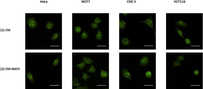
Fig. 11.

Representative images from immunofluorescence staining with anti-PARP1 antibody in HeLa, MCF7, CNE-2 and HCT116 cells after X-ray irradiation. IRCs were treated for 30 min with 15 mL of (1) CM and (2) CM + BAY5 post-irradiation. After the various treatments for 30 min, 15 mL of FM was used to replace the medium in the recipient dishes for 11.5 h (i.e. until 12 h post-irradiation) and immunofluorescent staining was performed. Scale bar = 25 μm.
