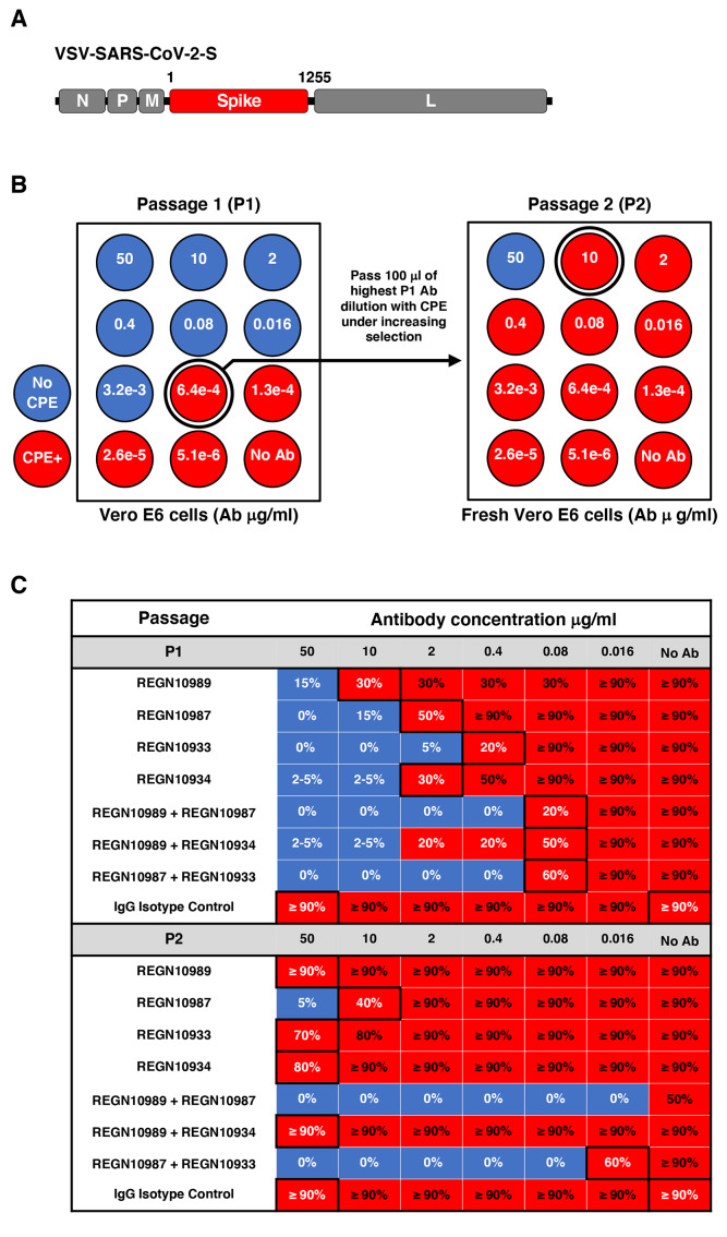
Fig. 1. Escape mutant screening protocol.
(A) A schematic is displayed of the VSV-SARS-CoV-2-S virus genome encoding residues 1-1255 of the spike protein in place of the VSV glycoprotein. N, nucleoprotein, P, phosphoprotein, M, matrix, and L, large polymerase. (B) A total of 1.5 × 10⁶ pfu of the parental VSV-SARS-CoV-2-S virus was passed in the presence of antibody dilutions for 4 days on Vero E6 cells. Cells were screened for virus replication by monitoring for virally induced cytopathic effect (CPE). Supernatants and cellular RNAs were collected from wells under the greatest antibody selection with detectable viral replication (circled wells; ≥20% CPE). For a second round of selection, 100uL of the P1 supernatant was expanded for 4 days under increasing antibody selection in fresh Vero E6 cells. RNA was collected from the well with the highest antibody concentration with detectable viral replication. The RNA was deep sequenced from both passages to determine the selection of mutations resulting in antibody escape. (C) The passaging results of the escape study are presented with the qualitative percentage of CPE observed in each dilution (red ≥ 20%CPE and blue < 20% CPE). Black boxes indicate dilutions that were passaged and sequenced in P1 or sequenced in P2. A no antibody control was sequenced from each passage to monitor for tissue culture adaptations.

