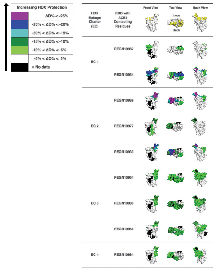
Fig. 3. HDX-MS determines mAb interaction on spike protein RBD.
3D surface models for the structure of the spike protein RBD domain showing the ACE2 interface and HDX-MS epitope mapping results. RBD residues that make contacts with ACE2 (21, 22) are indicated in yellow (top). RBD residues protected by anti–SARS-CoV2 spike antibodies are indicated with colors that represent the extent of protection, as determined by HDX-MS experiments. RBD residues in purple and blue indicate sites of lesser solvent exchange upon antibody binding that have greater likelihood to be antibody-binding residues. The RBD structure is reproduced from PDB 6M17 (21).

