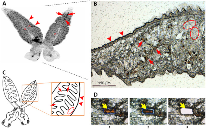
Fig 1. Laser microdissection of chosen tissues of E. nipponicum.
(A) An overview of E. nipponicum body. (B) Part of microdissected section. A branched intestine filled with host blood is clearly visible (red arrow). Tegument is visible at the edge of the section (red arrowhead). Parenchyma is the area with no visible organelles (red ovals). (C) A schematic drawing of worm anatomy and dissected areas. (D) Microdissection process: 1) selection of desired area; 2) laser section; 3) catapulting specimen to collection tube. Yellow arrows point to the dissected area.

