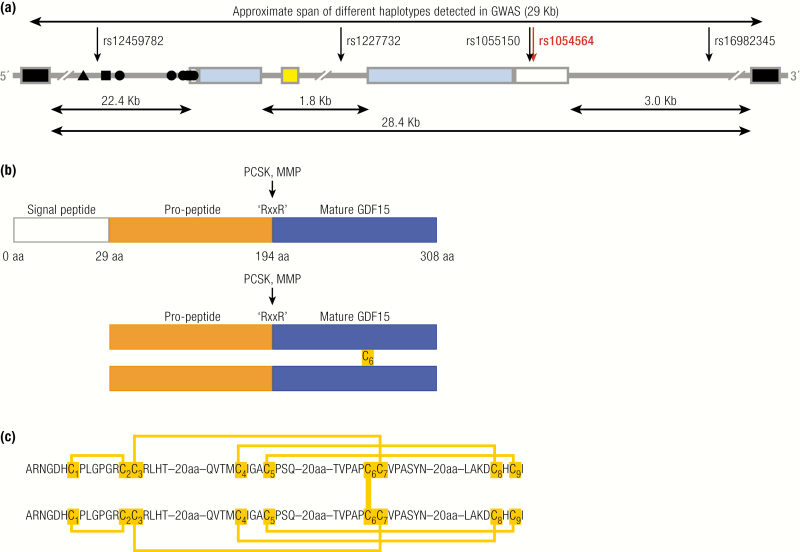
Figure 1.
GDF15: Gene and protein structure. A: Schematic representation of the human genomic segment containing GDF15 between neighbor genes PGPEP1 and LRCC25. GDF15 in blue (coding exons) and white (UTR); the 3’ and 5’ coding exons of PGPEP1 and LRCC25, respectively, in black; miRNA miR-3189 in yello; and the experimentally established transcription factor binding sites in the promoter for ▲ CHOP, ■ P53, and ● Sp1/Egr1. Sense of the GDF15 transcript is from left to right. The arrows indicate the putative causal SNPs in 5 different haplotypes that might influence GDF15 transcription. The red arrow indicates the rs1054564 variant in the GDF15 3’-UTR that has been experimentally validated to alter GDF15 expression via altered miRNA binding (16). B: Schematic demonstrating the signal peptide (blue), propeptide (orange), and mature peptide of GDF15. Pairing of the 6th cysteine in 2 pro-GDF15 monomers forms a pro-GDF15 dimer, which can be secreted and bound to the extracellular matrix or proteolytic cleavage at an “RXXR” motif and can liberate the mature peptide, which is secreted and circulates as a bioactive homodimer. C: Schema illustrating the cysteine–cysteine pairing within, and between, GDF15 monomers.

