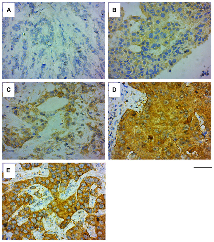
Figure 3. GRB7 expression and localization in invasive ductal carcinoma (IDC) of the breast.
Representative images of IDC of the breast tissues stained with the GRB7 antibody (brown) and Hematoxylin (blue). (A) IDC showing no GRB7 expression (0). (B) IDC showing weak cytoplasmic GRB7 expression (1+). (C) IDC showing medium cytoplasmic GRB7 expression (2+). (D) IDC showing strong cytoplasmic GRB7 expression (3+). (E) IDC showing strong cytoplasmic GRB7 expression with membranous accentuation (3+). Images were taken with a 40 × objective, scale bar = 50 μm.

