
Figure 2.
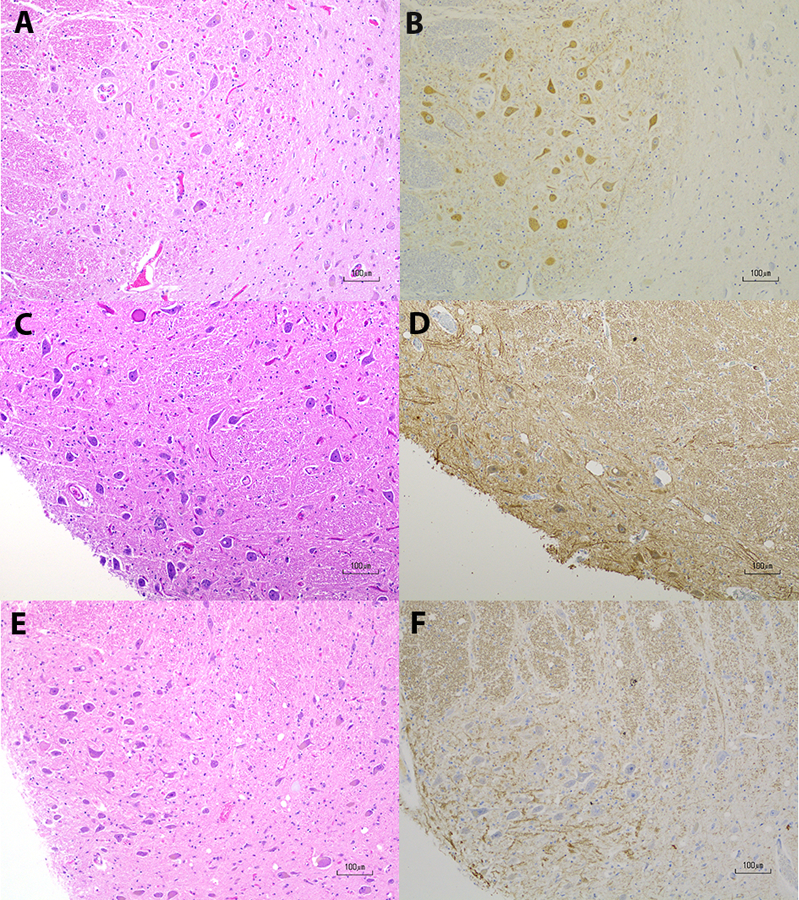
Choline Acetyltransferase Immunohistochemistry in the Pedunculopontine Nucleus
A & B. Control pedunculopontine nucleus (PPN) with hematoxylin and eosin (H&E) staining in A and choline acetyltransferase (ChAT) immunohistochemistry in B. There is positive ChAT immunoreactivity in B; C& D. CD PPN with H&E staining in C and ChAT staining in D. While background staining is slightly higher in and around the PPN in D, there is a population of neurons with weakly positive immunoreactivity in the cytoplasm, which is the expected pattern of ChAT staining; E & F. CD PPN with H&E staining in E and ChAT staining in F. F is an example of negative immunoreactivity in PPN neurons. Scale bars in A-F are 100 μm.