
Figure 2.
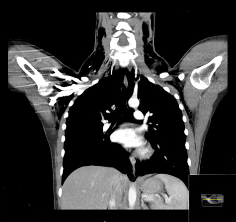
Coronal CT demonstrating extensive surgical emphysema in the neck, supraclavicular fossae, axillae and upper chest wall with moderate pneumomediastinum.
Official websites use .gov
A
.gov website belongs to an official
government organization in the United States.
Secure .gov websites use HTTPS
A lock (
) or https:// means you've safely
connected to the .gov website. Share sensitive
information only on official, secure websites.
Coronal CT demonstrating extensive surgical emphysema in the neck, supraclavicular fossae, axillae and upper chest wall with moderate pneumomediastinum.