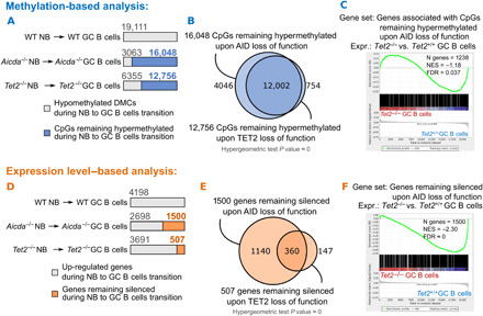
Fig. 5. TET2-deficient GC B cells manifest AID loss-of-function signature.

(A) Examination of 5mC accumulation during NB to GC B cell differentiation in TET2- and AID-deficient mouse models. CpGs (19,111) that are hypomethylated during normal NB to GC B cell differentiation in a WT mouse model were taken as a reference. (B) Venn diagram showing the overlap between CpGs that accumulate 5mC in Aicda−/− and in Tet2−/− NB to GC B cell differentiation. (C) GSEA enrichment analysis of 1238 genes accumulating 5mC during Aicda−/− NB to GC B cell transition, in Tet2−/− versus Tet2+/+ GC B cells. (D) Examination of gene inactivation during NB to GC B cell differentiation in TET2- and AID-deficient mouse models. Genes (4198) up-regulated during normal NB to GC B cell differentiation in a WT mouse model were taken as a reference. (E) Venn diagram showing the overlap between genes that remain silenced during Aicda−/− and Tet2−/− NB to GC B cell differentiation. (F) Enrichment of the 1500 genes that remain silenced during Aicda−/− NB to GC B cells transition, in Tet2−/− versus Tet2+/+ GC B cells.
