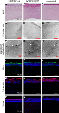
Fig. 3. Histopathology, TEM, and immunohistochemistry of the LiQD Cornea at 12 months.

(A to C) Paraffin-embedded sections of porcine cornea stained with hematoxylin and eosin (H&E) show multilayered, nonkeratinizing epithelia in all three samples. (D to F) TEM images of corneal epithelium in all three samples. (G to I) Epithelial cells showed abundance of desmosomes between cells (arrowheads). (J to L) Fully regenerated corneal tear film mucin stained with fluorescein isothiocyanate–conjugated lectin (green) from Ulex europaeus is seen in the LiQD Cornea. This is similar to the tear film in the controls. (M to O) Cytokeratin 12 (red), a marker for fully differentiated corneal epithelial cells, is present in the regenerated LiQD Cornea as in controls. (P to R) CD163 staining (red) shows that a few mononuclear cells are present in stroma of all three samples. Cell nuclei were stained blue with DAPI.
