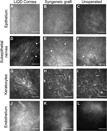
Fig. 5. In vivo confocal microscopy images of the LiQD Cornea compared to a healthy unoperated cornea and a syngeneic graft at 12 months after surgery.

Regenerated corneal epithelial cells cover the surface of the LiQD Cornea (A) as with the syngeneic graft (B) and untreated cornea (C). Regenerated nerves (arrowheads) were found at the sub-basal epithelium within the LiQD Cornea (D), ran parallel to one another, and were morphologically similar to those found in the unoperated cornea (F). Nerves in the syngeneic graft were less distinct (E). Keratocytes were present in all corneas (G to I). The unoperated endothelium remained intact and healthy in all corneas (J to L). Scale bars: 100 μm.
