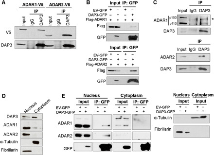
Fig. 1. DAP3 interacts with ADARs in the nucleus.

(A) Co-IP analysis of protein extracts from EC109 cells transfected with V5-tagged ADAR1 or ADAR2. Western blot (WB) analysis of DAP3-pulldown products was conducted using V5 and DAP3 antibodies. IgG, immunoglobulin G. (B) Co-IP analysis of protein extracts from EC109 cells cotransfected with the indicated tagged ADAR1/2 and DAP3 constructs, using a GFP-trap system. WB analysis of GFP-pulldown products was conducted using Flag and GFP antibodies. EV-GFP, GFP empty vector. (C) Co-IP analysis of protein extracts from EC109 cells. IP was performed with a DAP3 antibody, followed by WB analysis of DAP3-pulldown products using ADAR1, ADAR2, and DAP3 antibodies. *, nonspecific band. (D) WB analysis of the nuclear and cytoplasmic fractions of EC109 cells. (E) Co-IP analysis of the nuclear and cytoplasmic fractions of EC109 cells transfected with DAP3-GFP or EV-GFP. WB analysis of GFP-pulldown products was conducted using the indicated antibodies (left). α-Tubulin (cytoplasmic control) and fibrillarin (nucleic control) were analyzed in the input after fractionation (right). (A to C and E) One percent of the total cell lysate was loaded as an input control.
