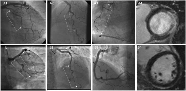
Figure 1.
Exemplar images of spontaneous coronary artery dissection patients with varying infarct mass. Patient A had three-vessel spontaneous coronary artery dissection (A1–A3) leading to a large lateral infarct but sparing of the inferior, septal, and anterior walls (A4). Patient B had two-vessel spontaneous coronary artery dissection involving the left anterior descending and circumflex coronaries (B1 and B2) but with no late gadolinium enhancement on cardiac magnetic resonance imaging (B4).

