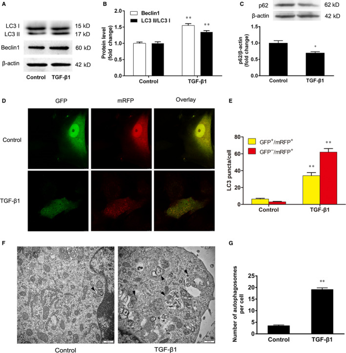
Figure 2.

The effect of TGF‐β1 on autophagy in VSMCs. The VSMCs were treated with TGF‐β1 (5 ng/mL) for 24 h. A, B, The conversion of LC3 I to LC3 II and the expression of Beclin1 were determined by Western blotting. C, The expression of p62 was determined by Western blotting. D, E, The VSMCs were infected with mRFP‐GFP‐LC3 adenovirus for 12 h, and then the cells were treated with TGF‐β1 for 24 h. The merged images (yellow) show overlay of GFP‐LC3 (green) and mRFP‐LC3 (red). The accumulation of red and yellow puncta were quantified for both control and TGF‐β‐treated cells. F, G, The autophagosome formation was analysed by transmission electron microscopy. Solid black arrowheads indicate the presence of autophagosomes. At least 20 cells were collected for statistical analysis in each group. The results are expressed as the mean ± SEM, n = 3. Statistical significance was determined using ANOVA by Student's t test. *P < 0.05 and **P < 0.01 vs. the control group
