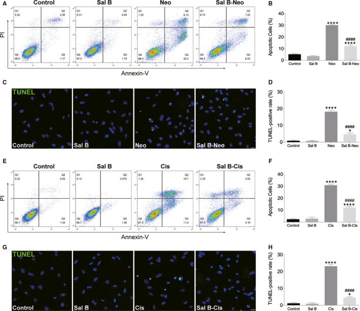
Figure 2.

Sal B reduced neomycin‐ and cisplatin‐induced apoptosis in HEI‐OC1 cells. (A and B) HEI‐OC1 cells were pre‐treated with or without Sal B for 2 hours, followed by exposure to 10 mM neomycin for 24 hours except those in the non‐treated control (control) group, and cell apoptosis was determined by flow cytometry using Annexin V and PI staining kit. (C and D) Apoptosis was assessed by TUNEL staining. (E and F) Effect of Sal B on cisplatin‐induced cell apoptosis. (G and H) Apoptosis was assessed by TUNEL staining. Representative images of TUNEL staining from different treatments (G) and quantification of the data (H) were shown. Data were presented as the mean ± SEM. *P < 0.05 and ****P < 0.0001 vs the non‐treated control group; #### P < 0.0001 vs the group treated with 10 mM neomycin or 30 μM cisplatin only. Scale bars = 20 μm
