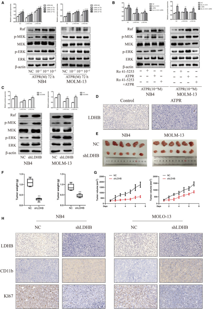
Figure 4.

ATPR regulates the Raf/MEK/ERK signalling pathway through LDHB and the effects of LDHB on tumour growth in vivo (A) Protein expression of Raf, p‐MEK, MEK, p‐ERK and ERK was determined by Western blotting analysis after treatment with ATPR for different concentration (10−7,10−6,10−5) (Mean ± SD, n = 3). (B) Protein expression of Raf, p‐MEK, MEK, p‐ERK and ERK was determined by western blotting analysis after treatment with ATPR in the absence or in the presence of the RARα‐selective antagonist Ro 41‐5253 (Mean ± SD, n = 3). (C) Protein expression of Raf, p‐MEK, MEK, p‐ERK and ERK was determined by Western blotting analysis after LDHB depletion in NB4 and MOLM‐13 cells (Mean ± SD, n = 3). (D) Representative two tumour tissues from vehicle control mice and ATPR‐treated mice group was fixed and immunohistochemistry staining for LDHB. (E, F) Tumour images and weights at experimental endpoints in NC and shLDHB xenografts (Mean ± SD, n = 6). (G) Tumour volumes were measured every day (Mean ± SD, n = 6). (H) Immunohistochemistry staining for LDHB, CD11b and KI67 of NC and shLDHB in NB4 and MOLM‐13 group. *P < 0.05, **P < 0.01 versus control group
