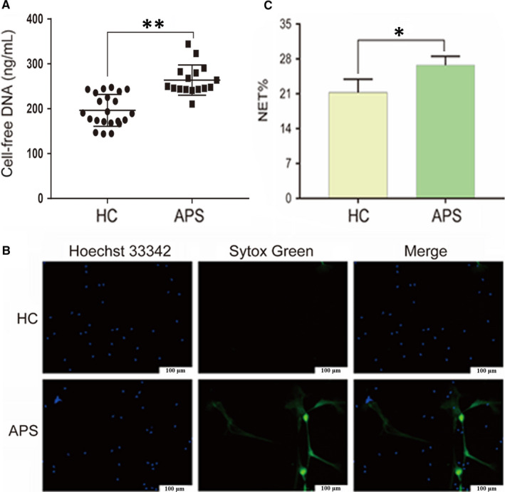
FIGURE 2.

Neutrophils from patients with APS are primed to NETosis. Neutrophils were freshly isolated from HCs (n = 22) or pregnant women with APS (n = 16) and incubated in 96‐well plates without any in vitro stimulation for 2 h. A, Cell‐free DNA levels in the supernatant. B, Representative live‐cell images of HC and APS neutrophils. Extracellular DNA was detected with the cell‐impermeable dye SYTOX Green. Intracellular DNA was detected with the cell‐permeable dye Hoechst 33342. Scale bars = 100 microns. C, NET release was scored as presented in panel B. *P < 0.05, **P < 0.01. Data are presented as the mean ± SD (A) or SEM (C; at least three independent experiments). APS, antiphospholipid syndrome; HC, healthy control; NET, Neutrophil extracellular trap
