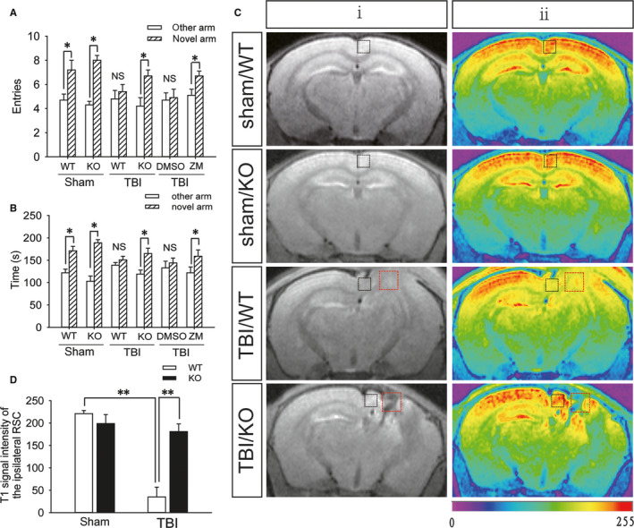
FIGURE 1.

A2AR knockout is beneficial to the function of the retrosplenial cortex and spatial recognition memory after TBI. A, The number of entries into the novel arm and other arm by mice in the sham (WT and KO), WT + TBI, KO + TBI, WT + TBI +DMSO and WT + TBI + ZM241385 groups; DMSO: dimethyl sulfoxide and ZM241385: A2AR antagonist. Data are presented as the means ± SDs, n = 15, novel arm vs other arm, *P < 0.05. B, Exploration time in the novel arm and other arm by mice from the sham (WT and KO), WT + TBI, KO + TBI, WT + TBI +DMSO and WT + TBI +ZM241385 groups. Data are presented as the means ± SDs, n = 15, novel arm vs other arm, *P < 0.05. C, T1‐weighted MEMRI of the brains of mice from the sham (WT and KO), WT + TBI and KO + TBI groups; the images in (ii) are colour‐coded versions of the images from (i) to better visualize the enhancement patterns. An increase in the red signal represents stronger neuronal activity. The black square frame indicates the ipsilateral retrosplenial cortex and the red square frame indicates the injured part. D, Quantification of the T1 signal intensity in the ipsilateral retrosplenial cortex shown in (C). Data are presented as the means ± SDs, n = 3, WT + TBI group vs KO + TBI group, **P < 0.01. KO, knockout; MEMRI, manganese‐enhanced magnetic resonance imaging; TBI, traumatic brain injury; WT, wild‐type
