
Figure 6: EGFR Site N420 Fucosylated Glycoform, EThcD MS/MS Spectrum of EGFR Glycopeptide 406TKQHGQFSLAVVSLNITSLGLR427 + HexNAc4Hex5dHex2 from HSC-3 Cells After Treatment with ICG-001.
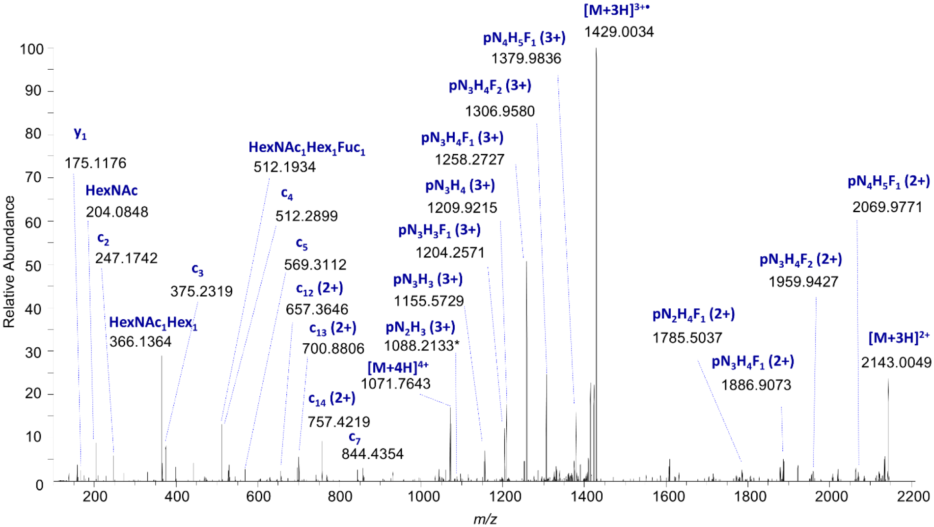
Electron-transfer/higher-energy collision dissociation (EThcD) mass spectrum of EGFR Site N420 glycopeptide, [M+4H]4+ m/z 1071.7526, demonstrating evidence of N-glycan “outer arm” fucosylation; the presence of the HexNAc+Hex+dHex oxonium ion at m/z 512.1934, and the lack of dHex shift associated with fragments that contain only the trimannosyl chitobiose core (pN2H3), suggest that both fucose residues are located on the outer arms, consistent with expression level data. p = peptide, N = HexNAc, H = hexose, F = dHex.