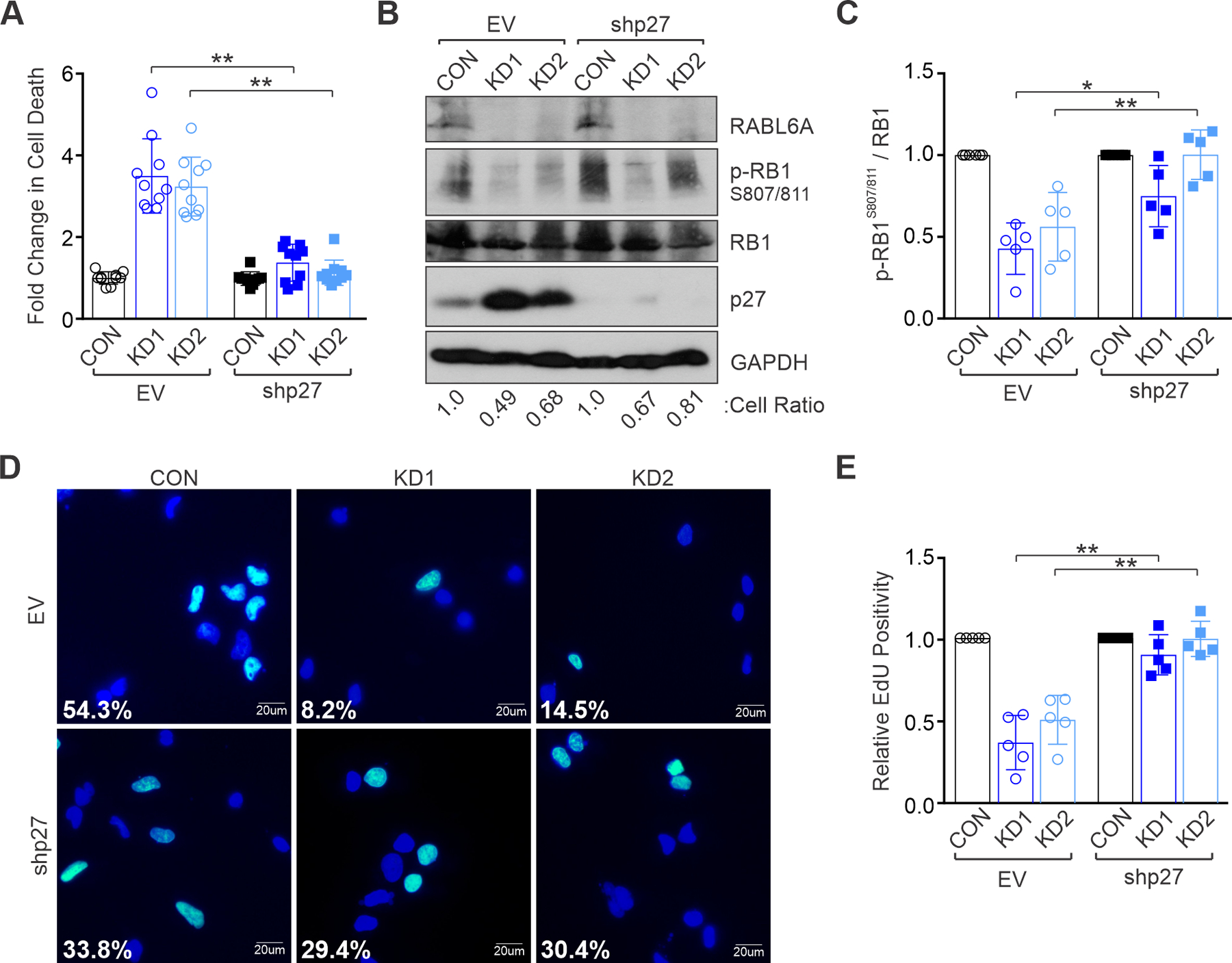
Figure 4. RABL6A promotes MPNST cell proliferation and survival via p27-RB1 inactivation.

S462 MPNST cells were transduced with empty vector (EV) or p27 shRNA viruses and subsequently infected with control (CON) or RABL6A shRNAs (KD1 and KD2). (A) Quantification of the fold change in cell death after RABL6A knockdown shows that p27 inactivation rescues cell death caused by RABL6A loss. (B) Representative westerns show partial (KD1) to full (KD2) restoration of RB1 phosphorylation (p-RB1) upon co-depletion of p27 with RABL6A. Below, cell ratio (relative to control cells) for the displayed experiment. (C) ImageJ quantification of western data from three or more experiments showing partial to full restoration of phospho-RB1 when p27 is depleted in tandem with RABL6A. (D) Representative images of EdU staining show p27 inactivation rescues DNA synthesis in RABL6A depleted cells. Percent EdU positive cells (cyan colored nuclei versus dark blue nuclei for EdU negative cells) are denoted. (E) Quantification of EdU positivity from three independent experiments. Data in A, C, and E are from three of more independent experiments. Error bars, SD from mean. P-value, Student’s t-test with Bonferroni’s correction. *, p<0.05; **, p<0.01.
