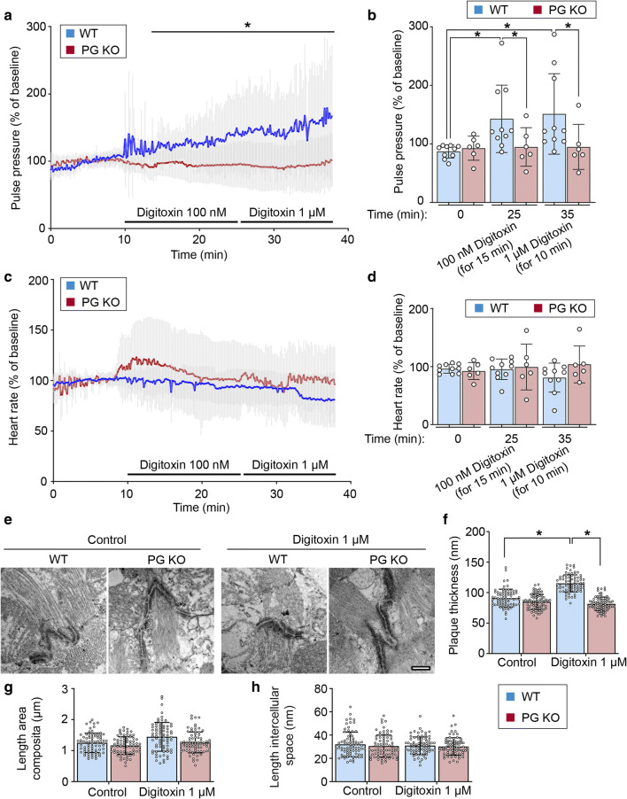
Fig. 1.
Positive inotropy induced by digitoxin is dependent on plakoglobin (PG) in ex vivo-perfused hearts. Mean pulse pressure (a) and heart rate (c) curve of hearts of 12-week-old WT or PG KO mice perfused with indicated digitoxin concentrations in an ex vivo Langendorff setting. Lines indicate mean values ± SD. Two-tailed, unpaired Student’s t test, *P < 0.05 vs. WT. b, d Represent corresponding values of N = 10 mouse hearts for WT and 6 mouse hearts for PG KO at indicated time points. Every dot corresponds to one individual heart, mean ± SD. Two-way ANOVA with Tukey’s post-hoc test. e Representative transmission electron microscopy images from cardiac slices derived from hearts of 12-week-old WT or PG KO mice and treated with digitoxin 1 µM for 60 min. Scale bar: 375 nm. N = 3 mice per condition. Analysis of junctional plaque thickness (f), length of area composita junctions (g), and length of intercellular space (h) corresponding to e. Every dot corresponds to one ICD, mean ± SD. Two-way ANOVA with Tukey’s post-hoc test

