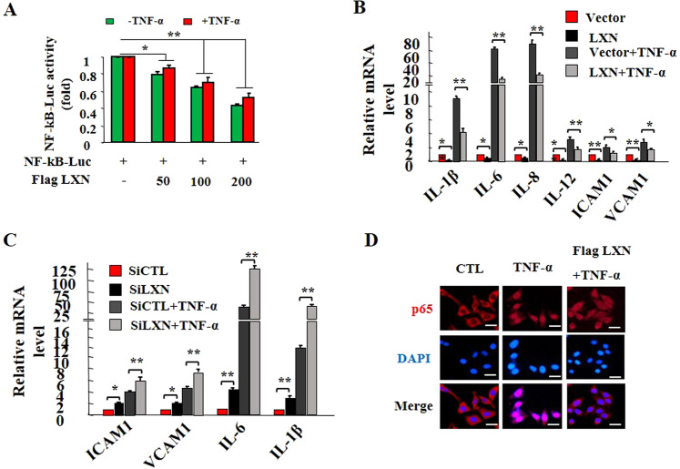
Figure 2.
LXN attenuates inflammatory response in HIECs. (A) HEK293T cells cultured in 6-well plates were co-transfected with 200 ng NF-κB-Luc reporter plasmid and 0, 50 ng, 100 ng and 200 ng of Flag-LXN as indicated, and 50 ng RL-SV40 was used as a control. 48 h after transfection, cells were treated with TNF-α (20 ng/mL) for 6 h, and then cells were lysed, and the reporter activity was determined with a luminescence counter using the Dual-Luciferase Reporter Assay System. (B) HIEC cells were seeded in 6-well plates 48 h after transfection of Flag-LXN or control plasmid, the cells were stimulated with TNF-α (20 ng/mL) for 6 h, and the expression of IL-1β, IL-6, IL-8, IL-12, ICAM1 and VCAM1 transcripts was examined by qPCR. (C) HIEC cells were treated with LXN siRNA for 72 h, the cells were stimulated with TNF-α (20 ng/mL) for 6 h, and the expression of IL-1β, IL-6, ICAM1 and VCAM1 transcripts was examined by qPCR. (D) HIEC cells were seeded in 6-well plates 48 h after transfection of Flag-LXN or control plasmid, the cells were stimulated with TNF-α (20 ng/mL) for 30 min, the cells were then washed, fixed, and stained with anti-p65 antibody. Scale bars = 10 μm. Data are representative of three independent experiments. *p < 0.05; **p < 0.01.

