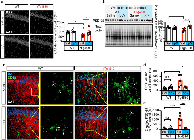
Fig. 7.
PTEN inhibition prevents microglia mediated cell and synapse loss in rTg4510 mice. a Surface rendering of DAPI from all groups, showing a rescue in cellular density of the cellular layer of the CA1 region of the hippocampus following chronic PTEN inhibition. b Representative western blots and quantification for PSD-95 and total protein in brain lysates from saline- or bpV-treated rTg4510 and WT mice.), revealing increased PSD-95 levels in bpV- compared to saline-treated rTg4510 mice. c 3D renderings of PSD-95 (red), CD68 (green) and DAPI (blue) in the hippocampus of saline- or bpV-treated rTg4510 and WT mice; arrowheads indicate engulfment. Scale bar in composite images: 20 μm; in zoomed-in images: 5 μm. d Quantification of CD68 volume, indicating that there is significantly less CD68 in rTg4510 mice treated with the PTEN inhibitor bpV. e Quantification of PSD-95 engulfed by microglia indicates that the rescue of PSD-95 loss in the bpV-treated group is due to a decrease in microglial engulfment. Data presented as mean ± SEM, *p < 0.05, **p < 0.01, ***p < 0.001, ****p < 0.0001; a, b, d, and e two-way ANOVA with Tukey’s multiple comparison test

