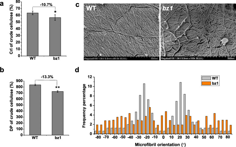
Fig. 4.
Detection of cellulose structural features. a Lignocellulose CrI of mature stems using the X-ray diffraction (XRD) method. b Cellulose DP of mature stems using the viscometry method. c Cryogenic scanning electron microscopy (Cryo-SEM) views of cellulose macrofibrils/microfibrils of the stem at booting stage after treatment with Updegraff reagent. d Distribution of macrofibrils/microfibrils orientation. The orientation of macrofibril/microfibril is represented as percentage frequency of the orientation of macrofibril/microfibril segments identified using the software Gwyddion (n = 3000 snakes from three images of three cells of three individual plants)

