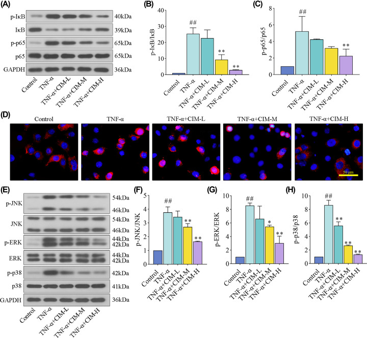
Figure 5. Cimifugin suppressed TNF-α-induced activation of NF-кB and MAPK signaling pathways in HaCaT cells.
(A–C) The expression levels of p-IкB, IкB, p-p65, and p65 proteins for NF-кB signaling pathway were detected by western blot. (D) The activation of p65 was evaluated using immnofluorescence staining. (E–H) The expression levels of p-JNK, JNK, p-ERK, ERK, p-p38, and p38 proteins for MAPK signaling pathway were tested by western blot. CIM-L, low dose of cimifugin; CIM-M, medium dose of cimifugin; CIM-H, high dose of cimifugin. ## P<0.01, compared with Control cells; * P<0.05, ** P<0.01, compared with TNF-α-treated cells.

