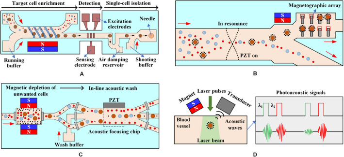
Figure 10.

The microfluidic system combines several external forces. A, The lateral magnetophoretic microseparator is fabricated using a bottom glass substrate with an inlaid ferromagnetic Permalloy wire array positioned at an angle of 5.7° to the direction of the flow. The microdispenser, including the impedance cytometer and the microshooter, is developed using a glass substrate with patterned gold electrodes. 112 B, Cells are acoustically focused to the center of the microchannel. Magnetically labeled cells are then deflected to the nonresonant portion of the microchannel via a gradient magnetic field. Finally, an array of micromagnets locally attracts magnetically labeled cells into microwells for on‐chip staining and analysis. 113 C, The blood sample passes through magnetic depletion that removes > 98% of unwanted blood cells, followed by an in‐line acoustic focusing and washing step, which removes debris and concentrates the sample prior to cell sorting. 114 D, CTCs targeted by two‐color nanoparticles can be illuminated by laser pulses at wavelengths of 639 and 900 nm with a delay of 10 μs. The laser beam is delivered either close to the external magnet or through a hole in the magnet by a fiber‐based delivery system 115
