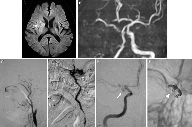
Fig. 1.
Patient imaging findings. (A) Diffusion-weighted magnetic resonance image shows hyper-intensity lesion in the right middle cerebral artery territory. (B) Magnetic resonance angiography shows occlusion of the right internal carotid artery (ICA). (C) The right ICA angiogram shows occlusion at the ICA origin. (D) Injection from the 9-Fr guiding catheter (GC) demonstrates massive extravasation from the terminal ICA. The origin of the ICA is wedged by the GC. (E) The delayed phase of the right ICA angiogram shows aneurysm-like dilatation at the terminal ICA (arrow). (F) The plain skull craniogram after parent artery occlusion shows a coil mass distributing at the terminal ICA. Some coil loop is distributed into the aneurysmal-like dilatation (arrow-head).

