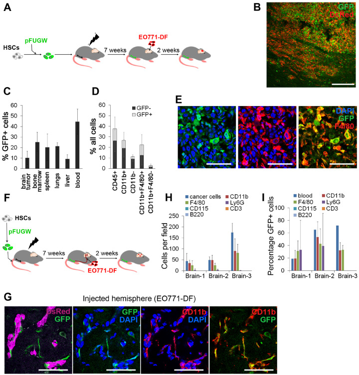
Figure 2.
Genetically modified hematopoietic stem cell (HSC) progeny in the context of macro- and micrometastases in the brain. A) Murine HSCs transduced with lentiviral pFUGW vector were transplanted into lethally irradiated recipient mice, and tumors were generated by intracranial implantation of EO771-DF cells. B) Detection of pFUGW-transduced HSC progeny (green fluorescent protein [GFP]+) in intracranial tumors generated according to the scheme in (A) by immunofluorescence (n = 7). Scale bar = 200 µm. C) Quantification of pFUGW-transduced HSC progeny (GFP+) in different tissues by flow cytometry; n = 3. D) Quantification of immune cell populations within EO771 intracranial tumors by flow cytometry. Total percentage of individual cell populations, as well as the proportion of cells derived from pFUGW-transduced HSCs is shown; n = 7. E) Immunofluorescence staining of intracranial EO771 tumors for macrophages/microglia (F4/80+) and GFP (n = 4). Scale bars = 50 µm. F) Murine HSCs transduced with lentiviral pFUGW vector were transplanted into lethally irradiated recipient mice, followed by administration of EO771-DF cells into the internal carotid artery. G) CD11b+GFP+ progeny of HSCs was observed in close association with EO771-DF micrometastases in the brain. Fluorescence images were obtained by confocal microscopy (n = 3). Scale bars = 50 µm. H) Cancer cells and micrometastases-associated cells belonging to different hematopoietic cell subpopulations were counted on immunofluorescence images; 4–8 micrometastases-containing coronal brain sections per animal (n = 3) were quantified. I) Percentages of GFP+ cells within micrometastases-associated hematopoietic cell populations were quantified using immunofluorescence images and overall percentages of GFP+ cells in matched blood by flow cytometry (group sizes as in H).

