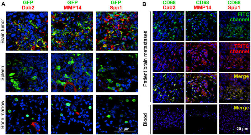
Figure 5.
Validation of expression of genes specific for brain metastases (BrM)-infiltrating myeloid cells in preclinical models and clinical specimens. A) The activity of gene promoters in the progeny of pFUGW-transduced hematopoietic stem cells was assessed in BrM, the spleen, and bone marrow of EO771 tumor-bearing mice (intracranial implantation model) with chimeric green fluorescent protein (GFP)+ bone marrow by performing immunofluorescence staining for the respective proteins and GFP (n = 3). Colocalization of DAB2, MMP14, and SPP1, respectively, with GFP is shown in merge images. Nuclei are stained with DAPI. Scale bars = 50 µm. B) Human BrM tissue and donor-matched blood were costained for the macrophage marker CD68 and for DAB2, MMP14, and SPP1, respectively (n = 4). Three further brain metastases specimens with donor-matched blood samples are shown in Supplementary Figure S6 (available online). Nuclei are stained with DAPI. Scale bars = 20 µm.

